Sodium sesquicarbonate elicits oxidative stress in erythrocytes, liver and kidney tissues.
Main Article Content
Abstract
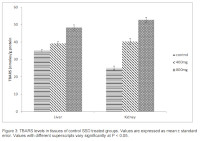
Sodium sesquicarbonate also known as Sodium sesquicarbonate dihydrate (SSD) has been used globally for centuries in food and traditional medical practices. There is paucity of scientific information on the safety of this common food additive. This study was designed to find out if the oral administration of SSD is capable of generating oxidative stress in erythrocytes, liver and kidney using albino rats as experimental models. The total number of animals used for this study was fifteen. The experimental animals were grouped into three. There were five animals in each group. The rats in the first group which was the control group, were dosed with 1 ml distilled water, while groups 2 and 3 were treated with 400 mg/kg and 800 mg/kg body weight (bw) of SSD, respectively, once daily per os for 28 days. After the duration of treatment, the erythrocytes, hepatic and kidney tissues were processed for the analysis. The biomarkers of oxidative stress, superoxide dismutase (SOD), catalase; and thiobarbituric acid reactive substances/ malondialdehyde (TBARS/MDA) were assayed. The result indicated that catalase enzyme activity was overexpressed in the red blood cells, liver and kidneys of the group that consumed the lower dose of SSD. The dose-dependent increase in the lipid peroxidation of the tissues as indicated by increased levels of MDA in the erythrocytes and TBARS in the tissues of the treated groups was significant (P < 0.05). The SOD enzyme activity in all the tissues assayed showed a dose-dependent decrease, which was significant at the probability level of 0.05. The consumption of SSD therefore caused lipid peroxidation and reduction in activity of the antioxidants present in the tissues studied.
Downloads
Article Details

This work is licensed under a Creative Commons Attribution-NonCommercial-NoDerivatives 4.0 International License.
References
Aebi, H. E. (1982). Catalase. In H. U. Bergmeyer (Ed.), Methods of Enzymatic Analysis (3rd ed., pp. 273–286). VerlagChemie, Weinheim, Germany.
Ajayi, A. F. and Akhigbe, R. E. (2017). Antispermatogenic mechanism of trona is associated with lipid peroxidation but not testosterone suppression. Journal of Human Reproductive Sciences, 10(2): 124. https://doi.org/10.4103/JHRS.JHRS_104_16
Ajiboye, J. A., Erukainure, O. L., Olasehinde, T., Obode, O. C. and Tugbobo-Amisu, A. O. (2018). Protective potential of Tetrapleura
tetraptera against trona (kaun)-induced hepatic injury in rat models. Comparative Clinical Pathology, 27(3): 627–633.
https://doi.org/10.1007/s00580-018-2639-z
Alburaidi, B. S., Alsenaidy, A. M., Al Hasan, M., Siddiqi, N. J., Alrokayan, S. H., Odeibat, H. A., Abdulnasir, A. J. and Khan, H. A. (2022).
Comparative evaluation of cadmiuminduced oxidative stress in camel and bovine erythrocytes. Journal of King Saud University - Science, 34(2): 101772. https://doi.org/10.1016/J.JKSUS.2021.101772.
Boriskin, P., Deviatkin, A., Nikitin, A., Pavlova, O. and Toropovskiy, A. (2019). Relationship of catalase activity distribution in serum and tissues of small experimental animals. IOP Conf. Ser.: Earth Environ. Sci, 403: 12113.
Cichoz-Lach, H. and Michalak, A. (2014). Oxidative stress as a crucial factor in liver diseases. World Journal of Gastroenterology : WJG, 20(25): 8082. https://doi.org/10.3748/WJG.V20.I25.8082
Czerska, M., Mikołajewska, K., Zieliński, M., Gromadzińska, J. and Wąsowicz, W. (2015). Today’s oxidative stress markers. Medycyna Pracy, 66(3): 393–405.
D’Azy, C. B., Pereira, B., Chiambaretta, F. and Dutheil, F. (2016). Oxidative and antioxidative stress markers in chronic glaucoma: A systematic review and metaanalysis. PLOS ONE, 11(12): e0166915. https://doi.org/10.1371/JOURNAL.PONE.0166915
Dhalla, N., Temsah, R. and Netticadan, T. (2000). Role of oxidative stress in cardiovascular diseases. Journal of Hypertension, 18(6):
–673.
Ene-Obong, H. N., Sanusi, R. A., Udenta, E. A., Williams, I. O., Anigo, K. M., Chibuzo, E. C., Aliyu, H. M., Ekpe, O. O. and Davidson, G. I. (2013). Data collection and assessment of commonly consumed foods and recipes in six geo-political zones in Nigeria: Important for the development of a National Food Composition Database and Dietary Assessment. Food Chemistry, 140(3): 539–546.
Glorieux, C., Zamocky, M., Sandoval, J. M., Verrax, J. and Calderon, P. B. (2015). Regulation of catalase expression in healthy and cancerous cells. In Free Radical Biology and Medicine 87: 84–97.
Gurgul, E., Lortz, S., Tiedge, M., Jörns, A. and Lenzen, S. (2004). Mitochondrial catalase overexpression protects insulin-producing cells against toxicity of reactive oxygen species and proinflammatory cytokines. Diabetes, 53(9): 2271–2280.
Gwozdzinski, K., Pieniazek, A. and Gwozdzinski, L. (2021). Reactive Oxygen Species and Their Involvement in Red Blood Cell Damage in Chronic Kidney Disease. Oxidative Medicine and Cellular Longevity, 2016: 6639199. https://doi.org/10.1155/2021/6639199
Hadwan, M. H. (2018). Simple spectrophotometric assay for measuring catalase activity in biological tissues. BMC Biochemistry, 19(1): 1–8.
Imafidon, K. E., Egberanmwen, I. D. and Omoregie, I. P. (2016). Toxicological and biochemical investigations in rats administered “kaun” (trona) a natural food additive used in Nigeria. Journal of Nutrition & Intermediary Metabolism, 6: 22-25
Inal, M., Kanbak, G., Şen, S., Akyüz, F. and Sunal, E. (1999). Antioxidant status and lipid peroxidation in hemodialysis patients
undergoing erythropoietin and erythropoietin-vitamin E combined therapy. Free Radical Research, 31(3): 211–216.
Jadhav, S. H., Sarkar, S. N., Aggarwal, M. and Tripathi, H. C. (2007). Induction of oxidative stress in erythrocytes of male rats subchronically exposed to a mixture of eight metals found as groundwater contaminants in different parts of India. Archives of Environmental Contamination and Toxicology, 52(1): 145–151.
Kakkar, P., Das, B. and Viswanathan, P. N. (1984). A modified spectrophotometric assay of superoxide dismutase. Indian
Journal of Biochemistry & Biophysics, 21(2): 130–132.
Kisic, B., Miric, D., Dragojevic, I., Rasic, J. and Popovic, L. (2016). Role of myeloperoxidase in patients with chronic kidney disease. Oxidative Medicine and Cellular Longevity, 2016: 1069743. https://doi.org/10.1155/2016/1069743
Kobayashi, M., Sugiyama, H., Wang, D., Toda, N., Maeshima, Y., Yamasaki, Y., Masuoka, N., Yamada, M., Kira, S. and Makino, H. (2005). Catalase deficiency renders remnant kidneys more susceptible to oxidant tissue injury and renal fibrosis in mice. Kidney International, 68(3): 1018–1031.
Kurian, G. A., Rajagopal, R., Vedantham, S. and Rajesh, M. (2016). The role of oxidative stress in myocardial ischemia and reperfusion injury and remodeling: revisited. Oxidative Medicine and Cellular Longevity,2016: 1656450. https://doi.org/10.1155/2016/1656450
Lewis, R. J. (2007). Hawley’s Condensed Chemical Dictionary. Journal of the American Chemical Society, 129(16):5296–5296.
Madesh, M. and Balasubramanian, K. A. (1998). Microtiter plate assay for superoxide dismutase using MTT reduction by superoxide. Indian Journal of Biochemistry & Biophysics, 35(3): 184–188.
Maurya, P. K., Kumar, P. and Chandra, P. (2015). Biomarkers of oxidative stress in erythrocytes as a function of human age.
World Journal of Methodology, 5(4): 216. https://doi.org/10.5662/WJM.V5.I4.216
Nielsen, J. M. and Dahi, E. (2002). Fluoride exposure of East African consumers using alkaline salt deposits known as magadi (trona) as a food preparation aid. Food Additives and Contaminants, 19(8): 709–714.
Nilakantan, V., Spear, B. T. and Glauert, H. P. (1998). Liver-specific catalase expression in transgenic mice inhibits NF-kappaB activation and DNA synthesis induced by the peroxisome proliferator ciprofibrate. Carcinogenesis, 19(4): 631–637.
Ohkawa, H., Ohishi, N. and Yagi, K. (1979). Assay for lipid peroxides in animal tissues by thiobarbituric acid reaction. Analytical
Biochemistry, 95(2): 351–358.
Okoye, J. O., Oranefo, N. O. and Okoli, A. N. (2016). Comparative Evaluation of the Effects of Palm Bunch Ash and Trona on the
Liver of Albino Rats. African Journal of Cellular Pathology, 6: 21–27.
Olson, K. R., Gao, Y., DeLeon, E. R., Arif, M., Arif, F., Arora, N. and Straub, K. D. (2017). Catalase as a sulfide-sulfur oxidoreductase: An ancient (and modern?) regulator of reactive sulfur species (RSS). Redox Biology, 12: 325–339.
Revin, V. V., Gromova, N. V., Revina, E. S., Samonova, A. Y., Tychkov, A. Y., Bochkareva, S. S., Moskovkin, A. A. and Kuzmenko, T. P. (2019). The Influence of oxidative stress and natural antioxidants on morphometric parameters of red blood cells, the hemoglobin oxygen binding
capacity, and the activity of antioxidant enzymes. BioMed Research International, 2019: 2109269.
https://doi.org/10.1155/2019/2109269.
Shafiq-Ur-Rehman. (1984). Lead-induced regional lipid peroxidation in brain. Toxicology Letters, 21(3): 333–337.
Sindhu, R. K., Ehdaie, A., Farmand, F., Dhaliwal, K. K., Nguyen, T., Zhan, C. De, Roberts, C. K. and Vaziri, N. D. (2005). Expression of catalase and glutathione peroxidase in renal insufficiency. Biochimica et Biophysica Acta (BBA) - Molecular Cell Research, 1743(1–2): 86–92.
Singh, M., Sandhir, R. and Kiran, R. (2010). Oxidative stress induced by atrazine in rat erythrocytes: mitigating effect of vitamin E.
Toxicology Mechanisms and Methods, 20(3): 119–126.
Skrzep-Poloczek, B., Poloczek, J., Chełmecka, E., Dulska, A., Romuk, E., Idzik, M., Kazura, W., Nabrdalik, K., Gumprecht, J., Jochem,
J. and Stygar, D. M. (2020). The oxidative stress markers in the erythrocytes and heart muscle of obese rats: relate to a high-fat diet
but not to DJOS bariatric surgery. Antioxidants (Basel, Switzerland), 9(2): 183. https://doi.org/10.3390/ANTIOX9020183
Tsikas, D. (2017). Assessment of lipid peroxidation by measuring malondialdehyde (MDA) and relatives in biological samples: Analytical and biological challenges. Analytical Biochemistry, 524:13–30.
Vaziri, N. D., Dicus, M., Ho, N. D., BoroujerdiRad, L. and Sindhu, R. K. (2003). Oxidative stress and dysregulation of superoxide
dismutase and NADPH oxidase in renal insufficiency. Kidney International, 63(1):179–185.
Yao, C., Behring, J. B., Shao, D., Sverdlov, A. L., Whelan, S. A., Elezaby, A., Yin, X., Siwik, D. A., Seta, F., Costello, C. E., Cohen, R. A., Matsui, R., Colucci, W. S., McComb, M. E. and Bachschmid, M. M. (2015). Overexpression of catalase diminishes oxidative cysteine modifications of cardiac
Proteins. PLOS ONE, 10(12): e0144025. https://doi.org/10.1371/JOURNAL.PONE.0144025
Yoshikawa, T. and Naito, Y. (2002). What Is oxidativestress? JMAJ, 45(7): 271-276.
Younus, H. (2018). Therapeutic potentials of superoxide dismutase. International Journal of Health Sciences, 12(3): 88.
/pmc/articles/PMC5969776/.
Zhang, P., Li, T., Wu, X., Nice, E. C., Huang, C. and Zhang, Y. (2020). Oxidative stress and diabetes: antioxidative strategies. Frontiers of Medicine, 14(5): 583–600.


