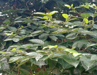
Characterization and antimicrobial evaluation of biomimetically synthesized silver nanoparticles using aqueous leaf extract of Morinda lucida Benth. (Rubiaceae)
Main Article Content
Abstract
Silver nanoparticles (AgNPs) are silver atom masses that are attracting widespread interest due to their diverse applications, particularly their activity as antimicrobial agents. Physico-chemical methods of AgNP synthesis are associated with high costs, high temperatures and toxic byproducts. Thus, the plant mediated pathway represents a better option. The indigenous medicinal plant Morinda lucida was employed in the fabrication of AgNPs. The nanoparticles were characterized using different analytical techniques and also evaluated for their antimicrobial potential. Phytochemical screening of the plant was also carried out. For the synthesis, 10 ml of aqueous M. lucida leaf extract was added to 90 ml of freshly prepared 3 mM silver nitrate (AgNO3) solution in a flask. The mixture was allowed to stand at ambient temperature, in a dark cupboard for 48 hours. Positive AgNP synthesis, indicated by a colour change from red to brown was further validated by UV-vis spectroscopy wherein an absorption peak at 460.51 nm was recorded. The utilitarian aspects of the particles were further characterized using scanning electron microscopy (SEM), fourier transform infrared (FTIR) spectroscopy, dynamic light scattering (DLS), X-ray diffraction (XRD) and energy dispersive X-ray spectroscopy (EDX). The SEM images showed that particles were round to irregular in shape. Amide, amine, alkene and alkynes were the most occurring functional groups from the FTIR spectra. Quantitative phytochemical analysis revealed the presence of flavonoids, terpenoids, phenols, reducing sugars and alkaloids in varying amounts, which play a significant role in the synthesis and stabilization of the AgNPs. The XRD diffractogram of AgNPs showed two peaks at 45.53° and 77.17° that correspond to miller indices of (200) and (311) respectively and an average crystalline size of 62.60 nm obtained using the Debye-Scherrer’s formula. The DLS result indicated a Z–average size of 235.1 nm and a polydispersity index (PDI) of 0.4. EDX analysis showed that elemental silver (Ag) had the highest atomic concentration of 64.50 %.
Using the agar well diffusion assay, the nanoparticles exhibited antimicrobial activity against P. aeruginosa (bacteria) and A. flavus (fungi). It can be concluded that M. lucida is capable of synthesizing stable, small-sized AgNPs with antimicrobial potential.
Downloads
Article Details

This work is licensed under a Creative Commons Attribution-NonCommercial-NoDerivatives 4.0 International License.
References
Addy, B. S., Owodo, H. T., Gyapong, R. N. K., Umeji, C. O. and Mintah, D. N. (2013).Phytochemical screening andantimicrobial study on the leaves of Morinda lucida (Rubiaceae). Journal of Natural Sciences Research, 3(14):131-135.
Adeleye, O. O., Ayeni, O. J. and Ajamu, M. A. (2018). Traditional and medicinal uses of Morinda lucida. Journal of Medicinal Plants Studies, 6(2): 249-254.
Ahmed, R. G., Hassan, S., Pal, S., Hashim, K. M., Sulaiman, R. H. and Khan, J. (2021). Antimicrobial efficacy of biogenic silver and zinc nanocrystals/nanoparticles to combat the drug resistance in human pathogens. In: Mallik, A. K (Ed.).Materials at the Nanoscale. IntechOpen, London, UK. pp. 184.
Ajayi, E. O., Odeyemi, S. W., Otunola, G. A. and Afolayan, A. J. (2017). Characterization and biological activities of silver nanoparticles synthesized from stems of Sarcocephalus latifolius (Sm) E. A. Bruce and Massularia acuminata (G. Don) Bullock ex Hoyl. (Rubiaceae). Asian
Journal of Chemistry, 29(6): 1240-1248.
Ajitha, B., Reddy, Y. A. K. and Reddy, P. S. (2015). Biosynthesis of silver nanoparticles using Momordica charantia leaf broth: Evaluation of their innate antimicrobial and catalytic activities, Journal of Photochemistry and Photobiology B: Biology, 146:1–9.
Alanazi, F. K., Radwan, A. A. and Alsarra, I. A. (2010). Biopharmaceutical applications of nanogold. Saudi Pharmeutical Journal, 18: 179-193.
Almadiy, A. A., Nenaah, G. E. and Shawer, D. M. (2018). Facile synthesis of silver nanoparticles using harmala alkaloids and
their insecticidal and growth inhibitory activities against the khapra beetle. Journal of Pest Science, 91(2): 727-737.
Association of Official Analytical Chemists (AOAC). (2005). Official Method of Analysis. 18th Ed. Association of Official Analytical Chemists International, Maryland, USA. Pp. 96
Dakal, T. C., Kumar, A., Majumdar, R. S., and Yadav, V. (2016). Mechanistic basis of antimicrobial actions of silver nanoparticles. Frontiers in Microbiology, doi.org/10.3389/fmicb.2016.01831.
Entsar, H. T. and Noha, M. A. (2016). Effect of silver nanoparticles on the mortality pathogenicity and reproductivity of entomopathogenic
nematodes. International Journal of Zoological Research, 12(3-4): 47-50.
Fissan, H., Ristig, S., Kaminski, H., Asbach, C. and Epple, M. (2014). Comparison of different characterization methods for nanoparticle dispersions before and after aerosolization. Analytical Methods, 6: 7324-7334.
Ge, L., Li, Q., Wang, M., Ouyang, J., Li, X. and Xing, M. M. Q (2014). Nanosilver particles in medical applications: Bio-Research Vol.21 No.3 pp.2065-2078 (2023) Synthesis, performance, and toxicity. International Journal of Nanomedicine, 9:2399-2407.
Geiss, O., Cascio, C., Gilliland, D., Franchini, F., and Barrero-Moreno, J. (2013). Size and mass determination of silver nanoparticles in an aqueous matrix using asymmetric flow field flow fractionation coupled to inductively coupled plasma mass spectrometer and ultraviolet–visible detectors, Journal of Chromatography A, 1321(38): 100-108.
Intertek (2018). Energy dispersive X-Ray (EDX) composition analysis. Available online: https://www.intertek.com/analysis/micro scopy/edx/. Accessed on 14th August, 2022.
Iravani, S., Korbekandi, H., Mirmohammadi, S. V. and Zolfaghari, B. (2014). Synthesis of silver nanoparticles: Chemical, physical and
biological methods. Research in Pharmaceutical Sciences, 9(6):385-406.
Jagtap, U. B. and Bapat, V. A. (2013). Green synthesis of silver nanoparticles using Artocarpus heterophyllus Lam. seed extract and its antibacterial activity. Industrial Crops and Products, 46: 132- 137.
Jalab, J., Abdelwahed, W., Kitaz, A. and Al-Kayali, R. (2021). Green synthesis of silver nanoparticles using aqueous extract of Acacia cyanophylla and its antibacterial activity. Heliyon, doi: 10.1016/j.heliyon.2021.e08033. Jesse, J. T. and Jesvin, S. (2018). A plausible antibacterial green synthesized AgNPs from Tridax procumbens leaf-flower extract, Journal of Pure and AppliedMicrobiology, 12(4):2135-2142.
Joseph, S. and Mathew, B. (2014). Microwave assisted biosynthesis of silvernanoparticles using the rhizomeextract of Alpinia galanga and evaluationof their catalytic and antimicrobialactivities. Journal of Nanoparticles, doi:http://dx.doi.org/10.1155/2014/967802
Kapil, A. (2005). The challenge of antibiotic resistance: Need to contemplate. Indian Journal of Medical Research, 121: 83-91.
Kora, A. J., Sashidhar, R. B. and Arunachalam, J. (2010). Gum kondagogu (Cochlospermum gossypium): A template for the green synthesis and stabilization of silver nanoparticles with antibacterial application. Carbohydrate Polymers, 82: 670-679.
Kelly, K. L., Coronando, E., Zhao, L. L. and Schatz, G. C. (2002). The optical properties of metal nanoparticles: The influence of size, shape and dielectric environment. Journal of Physical Chemistry B, 107:668–677.
Lin, P. C., Lin, S., Wang, P. C. and Sridhar, R. (2014). Techniques for physicochemical characterization
of nanomaterials. Biotechnology Advances, 32: 711-726.
Loo, Y. Y., Rukayadi, Y., Nor-Khaizura, M-A-R, Kuan, C. H., Chieng, B. W., Nishibuchi, M. and Radu, S. (2018).
In vitro antimicrobial activity of green synthesized silver nanoparticles against selected gram-negative foodborne pathogens. Frontiers
in Microbiology, doi: 10.3389/fmicb.2018.01555.
Mai-Prochnow, A., Clauson, M., Hong, J. and Murphy, A. B. (2016). Gram positive and Gram-negative bacteria differ in their sensitivity to cold plasma. Scientific Reports, 6: https://doi.org/10.1038/srep38610.
Masum, M. I., Siddiqa, M., Ali, K., Zhang, Y., Abdallah, Y., Ibrahim, E., Qiu, W., Yan, C. and Li, B. (2019). Biosynthesis ofsilver nanoparticles using Phyllanthus emblica fruit extract and its antibacterial activity against rice brown stripe pathogen Acidovorax oryzae. Frontiers
in Microbiology, doi.10.3389/fmicb.2019.00820
Nanakoudis, A. (2019). EDX analysis with SEM: How does it work?.https://www.thermofisher.com/blog/materials/edx-analysis-with-sem-how-does-it-work/. Accessed on 11th July, 2022
Nanocomposix (2012). UV/VIS/IR Spectroscopy Analysis of Nanoparticles. Available online:http://50.87.149.212/sites/default/files/nanoComposix%20Guidelines%20for%20UVvis%0Analysis.pdf . Accessed on 3rd August, 2022.
Netala, V. R., Bethu, M. S., Pushpalatha, B. P.,Baki, V. B., Aishwarya, S., Rao, J. V and Tartte, V. (2016). Biogenesis of silver nanoparticles using endophytic fungus Pestalotiopsis microspora and evaluation of their antioxidant and anticancer activities, International
Bio-Research Vol.21 No.3 pp.2065-2078 (2023)Journal of Nanomedicine, 11: 5683–5696.
Ogundare, A. O. and Onifade, A. K. (2009). The antimicrobial activity of Morinda lucida leaf extract on Escherichia coli. Journal of Medicinal Plants Research, 3(4):319-323
Olawuwo, O. S., Famuyide, I. M. and McGaw, L. J. (2022). Antibacterial and antibiofilm activity of selected medicinal plant leaf
extracts against pathogens implicated in poultry diseases. Frontiers in Veterinary Science, doi: 10.3389/fvets.2022.820304
Ondari, E. N., Omwenga, S. G. andMadanagopal, N. (2019). Tridax procumbens amines have a potential to synthesize
silver nanoparticles. Research Square,doi:10.21203/rs.2.19559/v1.
Pal, S., Tak, Y. K., and Song, J. M. (2007). Does the antibacterial activity of silver nanoparticles depend on the shape of the nanoparticle? A study of the gram- negative bacterium Escherichia coli. Applied and Environmental Microbiology,73(6): 1712-1720.
Panigrahi, S., Kundu, S., Ghosh, S., Nath, S. and Pal, T. (2004). General method of synthesis for metal nanoparticles. Journal of Nanoparticle Research, 6(4): 411-414.
Patra, J. K. and Baek, K. H. (2014). Green nanobiotechnology: Factors affecting synthesis and characterization
techniques, Journal of Nanomaterials,doi: http://dx.doi.org/10.1155/2014/417305
Paulkumar, K., Gnanajobitha, G., Vanaja, M., Pavunraj, M. and Annadurai, G. (2017). Green synthesis of silver nanoparticle and silver-based chitosan bionanocomposite using stem extract of Saccharum officinarum and assessment of its antibacterial activity. Advances in Natural Sciences: Nanoscience and Nanotechnology,
doi:/article/10.1088/2043-6254/aa7232
Pleus, R. (2012). Nanotechnologies-Guidance on Physico-chemical Characterization of Engineered Nanoscale Materials for Toxicologic Assessment. ISO: Geneva, Switzerland. pp. 33.
Prabhu, S. and Poulose, E. K. (2012). Silver nanoparticles mechanism of antimicrobial action, synthesis, medical applications, and toxicity effects. International Nano Letters, https://doi.org/10.1186/2228-5326-2-32
Reddy, N. V., Satyanarayana, B. M., Sivasankar,S., Pragathi, D., Subbaiah, K. V. and Vijaya, T. (2020). Eco-friendly synthesis of silver nanoparticles using leaf extract of Flemingia wightiana: Spectral characterization, antioxidant and
anticancer activity studies. SN Applied Sciences, https://doi.org/10.1007/s42452-020-2702-7
Robin, T. M. (2009). Introduction to powder diffraction and its application to nanoscale and heterogeneous
materials. In: Pacheco, K. and Jones, W.(eds.). Nanotechnology in Undergraduate Education. OUP USA, pp. 75–86
Sahu, N., Soni, D., Chandrashekhar, B., Satpute, D. B., Saravanadevi, S., Sarangi, B. K. and Pandey, R. A. (2016). Synthesis of silver nanoparticles using flavonoids: Hesperidin, naringin and diosmin, and their antibacterial effects and cytotoxicity. International Nano Letters, 6: 173–181.
Salomoni, R., Léo, P. and Rodrigues, M. (2015). Antibacterial activity of silver nanoparticles (AgNPs) in Staphylococcus aureus and cytotoxicity effect in mammalian cells. In: Mendez-Vilas, A. (Ed.). The Battle against Microbial Pathogens: Basic Science, Technological Advances and Educational Programs. Formatex Research Centre, Spain. pp. 851- 857
Shaik, M. R., Khan, M., Kuniyi, M., Al-Warthan, A., Alkhathlan, H. Z., Siddiqui, M. R. H., Shaik, J. P., Ahamed, A., Mahmood, A., Khan, M. and Adil, S. F. (2018). Plantextract-assisted green synthesis of silver nanoparticles using Origanum vulgare L. extract and their microbicidal activities. Sustainability, 10(913): 1 – 14.
Sharma, G., Nam, J. S., Sharma, A. R. and Lee, S. S. (2018). Antimicrobial potential of silver nanoparticles synthesized using medicinal herb Coptidis rhizome. Molecules, 23(9): https://doi.org/10.3390/molecules23092
Shriniwas, P. P. and Subhash, T. K. (2017). Antioxidant, antibacterial and cytotoxic potential of silver nanoparticles synthesized using terpenes rich extract of Lantana camara L. leaves. Biochemistry and Biophysics Reports, 10:76-81.
Shehzad, A., Qureshi, M., Jabeen, S., Ahmad, R., Alabdalall, A. H., Aljafary, M. A. and AlSuhaimi, E. (2018). Synthesis,
characterization and antibacterial activity of silver nanoparticles using Rhazya stricta. PeerJ,
https://doi.org/10.7717/peerj.6086
Skandalis, N., Dimopoulou, A., Georgopoulou,A., Gallios, N., Papadopoulos, D., Tsipas,D., Theologidis, I., Michailidis, N. and Chatzinikolaidou, M. (2017). The effect of silver nanoparticles size, produced using plant extract from Arbutus unedo, on their antibacterial efficacy. Nanomaterials, 7(7):178-183.
Srikar, S., Giri, D., Pal, D., Mishra, P. and Upadhyay, S. (2016). Green synthesis of silver nanoparticles: A review. Green and Sustainable Chemistry, 6:34-56.
Sumathi, S. and Thomas, A. (2017). Eco-friendly and antibacterial finishes of organic
fabrics using herbal composite microencapsules. International Journal of Pharmacy and Biological Sciences, 8: 310–321.
Tak, Y. K., Pal, S., Naoghare, P. K., Rangasamy, S. and Song, J. M. (2015). Shapedependent skin penetration of silver
nanoparticles: Does it really matter? Scientific Reports, 5: 16908 – 16911.
Tariq, M., Mohammad, K. N., Ahmed, B., Siddiqui, M. A. and Lee, J. (2022). Biological synthesis of silver
nanoparticles and prospects in plant disease management. Molecules, 27(15): 4754-4781.
Waseda, Y., Matsubara, E. and Shinoda, K. (2011). X-ray Diffraction Crystallography: Introduction, Examples and Solved Problems. Springer Verlag, Berlin, Germany. pp. 310
Wiley, B., Sun, Y., Mayers, B. and Xia, Y. (2005). Shape-controlled synthesis of metal nanostructures: The case of silver. Chemistry, 11: 454–463.
Xia, Z. K., Ma, Q. H., Li, S. Y., Zhang, D. Q, Cong, L., Tian, Y. L. and Yang, R. Y. (2016). The antifungal effect of silver
nanoparticles on Trichosporon asahii. Journal of Microbiology, Immunology and Infection, 49:182–188.
Yao, H. and Kimura, K. (2007). Field emission scanning electron microscopy for structural characterization of 3D
gold nanoparticle super lattices. In: Méndez-Vilas, A. and Díaz, J.(eds.). Modern Research and Educational Topics in Microscopy. Formatex Research Centre, Badajoz, Spain. pp. 568–575.
Zasshi, Y. (2019). Nanosize particle analysis by dynamic light scattering (DLS). Journal of
the Pharmaceutical Society of Japan, 139(2):237-248.
Zhang, X., Liu, Z., Shen, W. and Gurunathan, S. (2016). Silver nanoparticles: Synthesis, characterization, properties, applications and therapeutic approaches. International Journal of Molecular Science, 17(9): 1501-1534.


